vwackclnor.org vwackclnor.org
  • Home
  • About
  • Events
  • Veterinarians
    • Education/further training
    • Membership SignUp
    • Member Login
  • Logo
  • LupoMove®
    • General Information
    • LupoMove® puppy
    • LupoMove® junior
    • LupoMove® youth
    • LupoMove® adult
    • LupoMove® senior
    • LupoMove® sports & works
  • Dog-Owner
    • Puppy Passport
    • Puppy Passport II
    • Puppy Passport II (Interview)
    • Early detection, pain-free movement
    • Left Hindquarter lameness for 1 year
    • Shoulder lameness in Rottweiler in the "LupoMove-Youth-Program"
    • Chronic pain: always prescribe gabapentin
  • Contact

Shoulder lameness in Rottweiler in the "LupoMove-Youth-Program"

Our patient was presented to us at the end of April with shoulder lameness in the front right; she was practically unable to put any weight at all on the leg. The day before, the dog had had nothing wrong. She had only been in the garden, and there was no obvious explanation.

The young Rottweiler bitch is participating in our study, and had been inconspicuous during the last check at the beginning of March 2020. She had developed very well in the period from the 12th week to the 7th month. Then, just before the first control of the bimonthly rhythm, the bitch suddenly went lame in the front right leg!
You can see this in the first video.

Video
Patient movement from 30.4.2020


The subsequent radiology showed an injury of the shoulder in the area of the tuberculum supraglenoidale (attachment site of the biceps tendon), this is the tip of the shoulder in the transition to the upper arm and is an important area for the movement of the shoulder limb. It looked as if the joint had splintered, which explained the sudden lameness.

X-ray image: injury of the shoulder


If we briefly recall the anatomy of a dog's shoulder limbs, we have on one hand the shoulder blade, which moves along the chest and is connected to the dog's body by a muscular connection. This is called synsarcosis and the pseudo-articulation is a thoracic (chest) - scapular (shoulder blade) functional (joint-like) connecting axis. The bow joint, i.e. the region of injury in this dog, is a true joint as defined in the textbook. It is basically a ball and socket joint, but in function like a roller joint, comparable to the toe joints.

Illustration: bow joint


The owner was told that the dog must be kept on a line at all times, and mainly because of the pain and inflammation, the patient was prescribed meloxicam. A control should take place a good four weeks later. At that time, surgery was not an option, as the joint was too damaged to insert a screw for stabilisation. It might be necessary to do this at a later date, depending on developments.

In the control in May 2020, she was less lame, the pain was reduced and the resilience of the limb was significantly better.

Video
Patient from end of May 2020


X-ray image: Radiology also showed the beginning of calcification and callus formation


At this point, the patient was treated with orthopaedic manual kinematic control, and sent home with a controlled movement plan. The convalescence was positive, but surgical intervention could still not be ruled out. Perhaps this would not be necessary.

The next check-up was scheduled for the end of July. In her daily life, the lameness had practically disappeared - only in violent and especially short tight turns was the limping sometimes apparent.

Video
Patient from end of July 2020


X-ray image: good healing of the fragmentation


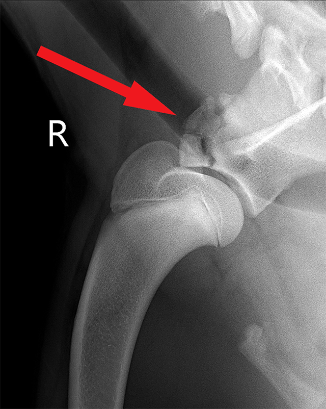
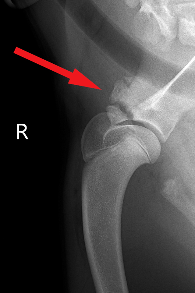
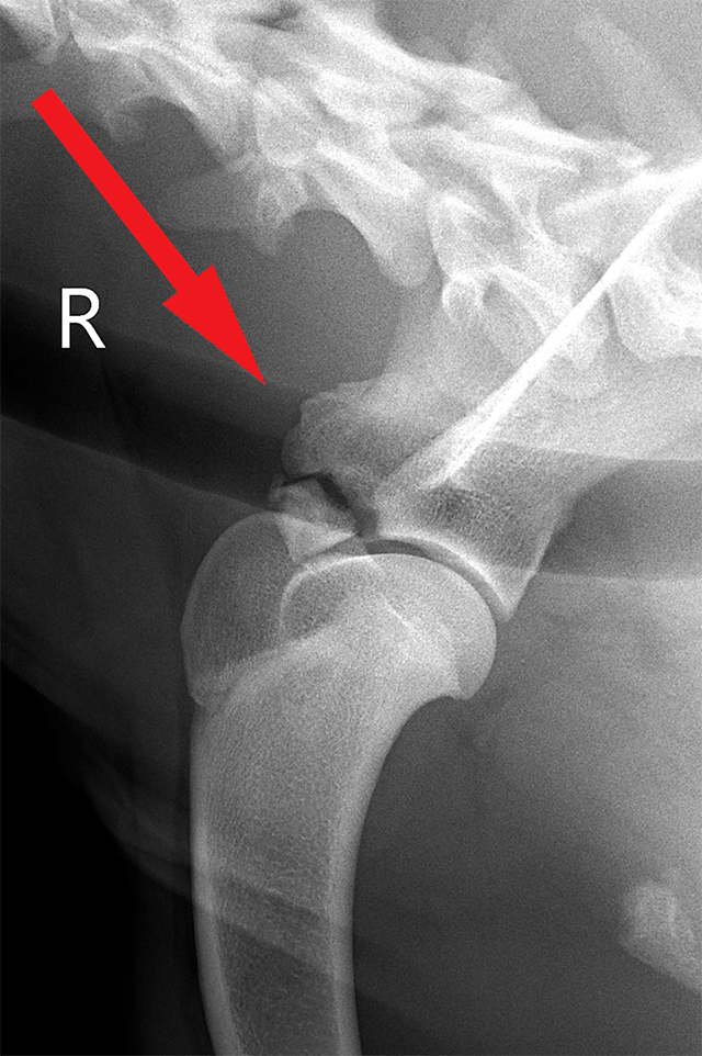
The X-ray study also showed a good healing of the fragmentation. Only the tubercle was not yet fully fused and mineralized. If this does not heal independently within the next 4 weeks, we could stabilise it with surgical intervention. The trend is quite good, however, and this should now heal on its own. In any case, we will keep an eye on it.

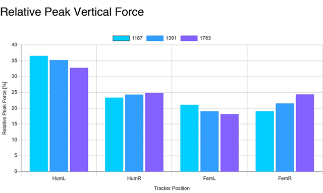
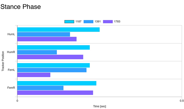
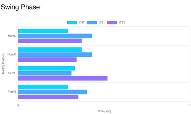
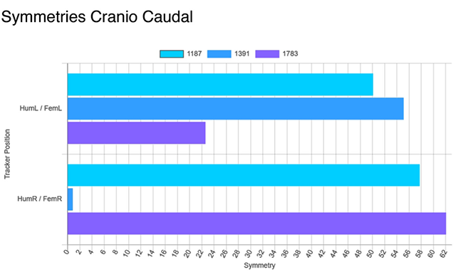
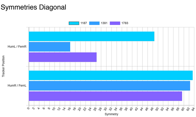
If we now examine this injury with the functional measurements, the development of lameness from the shoulder injury can be shown objectively and impressively.
For your information:

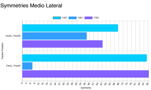
The light blue bars show the measurements at the end of April, the dark blue bars are from the end of May, and violet bars show the measurements from the end of July 2020, so you can always compare the same parameters. The best way to see the development of lameness is with the parameter "Relative Peak Vertical Force, PVF". Here you can see a slow rise of the bars at the right limb. This shows that the dog is able to put increasing weight on the diseased limb. The remaining parameters probably show a diseased right limb, but the patient tries to compensate for this, especially via the left pelvic limb. Nevertheless, at least we can see that the healing of the right shoulder limb is progressing.


In the next issue, I will introduce you to a young Rottweiler dog, 9 months, who had been treated with PTA for several months because of inflammation/pain in the growth joints in the forefoot joint. After a short examination, another diagnosis was found, which had drastic consequences. What was it? We’ll tell you next time.

www.vwackclnor.orginfo@vwackclnor.org